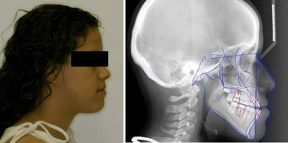


We have an active interventional service with over 40 breast procedures performed weekly.įellows have the responsibility of performing challenging procedures including localizations, ultrasound, and stereotactic-guided core biopsies and MRI biopsies each week. Our Breast MRI volume is robust with at least 20-25 cases per week. Screening services include mammography, MRI, and whole breast ultrasound. Our breast fellows have the opportunity to learn to interpret all types of imaging including screening and diagnostic mammograms, contrast-enhanced mammograms, breast ultrasound, and breast MRI. Our one-year, fellowship-level Breast Imaging curriculum provides an organized educational experience to graduate expert breast imaging consultants and practitioners for either the academic or private practice setting. On behalf of the Beth Israel Deaconess Medical Center (BIDMC) Breast Imaging Fellowship team, led by Janet Baum, MD, we thank you for your interest in our program. There have been studies on a great number of image processing applications carried out, such as OsiriX 3D®, Amira® or Analized®, which have definitely proven its efficacy in medical practice. It is also possible to interact with these 3D images simulating the surgery that will take place and producing predictions as to the postoperative outcome in soft and hard tissues. The move from 2D to 3D imaging provides surgeons, students and patients with extra information that cannot be obtained only from conventional tomographies. Anatomía Humana University of Valladolid Avda Ramón y Cajal, 7- Valladolid ABSTRACT volumetric data from Computed tomography (CT) or, more recently, Cone Beam Computed tomography (CBCT) can be converted into 3D images of craniofacial skeleton and the soft tissue covering it. Anatomía Humana University of Salamanca Avda.

They are extremely convenient, not only for preoperative planning of surgeries, but also as a communication tool withĭOLPHIN 3D: Technological Environment for Medical Image Processing on Training Alicia Hernández Salazar Servicio de Cirugía Oral y Maxilofacial Hospital Universitario de Salamanca Paseo San Vicente 88. Juanes Vázquez, Francisco PastorĭOLPHIN 3D: Technological Environment for Medical Image Processing on Training Alicia Hernández Salazar Servicio de Cirugía Oral y Maxilofacial Hospital Universitario de Salamanca Paseo San Vicente 88. Salazar, Alicia Hernández Méndez, Juan A. DOLPHIN 3D: technological environment for medical image processing on training DOLPHIN 3D: technological environment for medical image processing on training


 0 kommentar(er)
